Current Projects
Multiple sclerosis affects about 1 in 3,000 people and is a leading cause of disability in young people. While existing treatments prevent “flares”, or acute peripheral immune attacks, we have no options that substantially impact the insidious disease progression that occurs in many people or to repair and remyelinate existing lesions. Chronic, unresolving glial cell activation drives MS progression and is a common feature across neurodegenerative diseases. What do reactive glia do, and what triggers, perpetuates, or could reverse these states? How do immune cells in the brain’s parenchyma and borders (meninges) impact neurodegeneration? Current research is focused on these questions.
1. Tissue Resident Regulatory T cells and IL-33 in Neuroinflammation
Brain resident Tregs are protective
Regulatory T cells (Tregs) are a specialized subset of helper T cell whose play key roles in resolving inflammation and promoting wound healing. They’re defined by expression of the transcription factor FOXP3 and they arise either in the thymus or peripheral tissues. While scarce in the healthy brain, Tregs reside in the meninges and further accumulate during neuroinflammation. Among other markers, brain Tregs express high levels of the IL-33 receptor. Our lab is interested in how these tissue-resident Tregs are instructed by the CNS environment, and how they in turn shape reactive glial states in multiple sclerosis and its models.
IL-33 is a central nervous system (CNS) alarmin
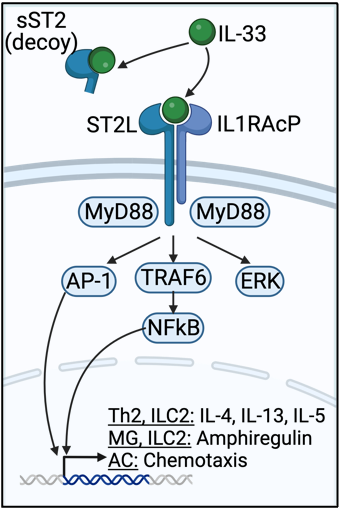
IL-33 is a nuclear cytokine expressed in the healthy CNS and released upon injury or stress by oligodendrocytes and astrocytes. Once released, IL-33 binds to its receptor, enhancing regulatory and type 2 inflammation through MyD88-dependent signaling in Th2 cells, Tregs, and type 2 innate lymphocytes (ILC2s). A decoy form of the receptor, sST2, inhibits IL-33 signaling. Interestingly, IL-33 is elevated in the CNS, cerebrospinal fluid (CSF), and plasma of people with multiple sclerosis, and appears to be beneficial; plasma IL-33 correlates with decreased multiple sclerosis lesion burden. IL-33 is similarly increased in mice with experimental autoimmune encephalomyelitis (EAE) and in the corpus callosum of mice fed the demyelinating toxin cuprizone (CPZ), and appears to promote recovery in those models. While IL-33 promotes recovery in the brain, the underlying beneficial mechanisms in the CNS remain poorly understood. We are studying this pathway using single cell RNA sequencing, animal models, and novel pharmacologics.
Garton T, Gadani SP, Gill AJ, Calabresi PA. Neurodegeneration and demyelination in multiple sclerosis. Neuron. 2024.
Gadani SP, Smirnov I, Smith AT, Overall CC, Kipnis J. Characterization of meningeal type 2 innate lymphocytes and their response to CNS injury. Journal of Experimental Medicine. 2017;214(2):285–296.
Gadani SP, Walsh JT, Smirnov I, Zheng J, Kipnis J. The glia-derived alarmin IL-33 orchestrates the immune response and promotes recovery following CNS injury. Neuron. 2015;85(4):703–709.
2. Use spatial transcriptomics to discover pathways driving grey matter pathology in multiple sclerosis
Grey matter pathology correlates with disability in progressive multiple sclerosis
Neuropsychiatric symptoms in multiple sclerosis are prevalent, with 60% of patients having cognitive impairment and 30% having depression, and disabling. While typically known as a disease of white matter, early histologic studies found focal areas of demyelination, neuron death, and microglia activation, as well overall atrophy of the cortical grey matter in multiple sclerosis. The extent of grey matter involvement closely correlates with cognitive impairment, fatigue, and depression, but modern multiple sclerosis therapies do not target this aspect of the disease. The underlying pathologic mechanism of cortical atrophy remains unknown, critically limiting our ability to design rational therapies that prevent it.
Mace JW*, Gadani SP*, Smith MD, Galleguillos D, Kang BG, Roy M, Liu M, Summers B, Garton T, Gharagozloo M, Gill AJ, Pardo CA, Sotirchos ES, Dawson VL, Dawson TM, Calabresi PA. Autoimmune neuroinflammation leads to neuronal death via MIF nuclease-mediated parthanatos. Nature Neuroscience. 2026;29:796–809. * Co-first author.
Inflammation in the subarachnoid space spatially relates to GMP
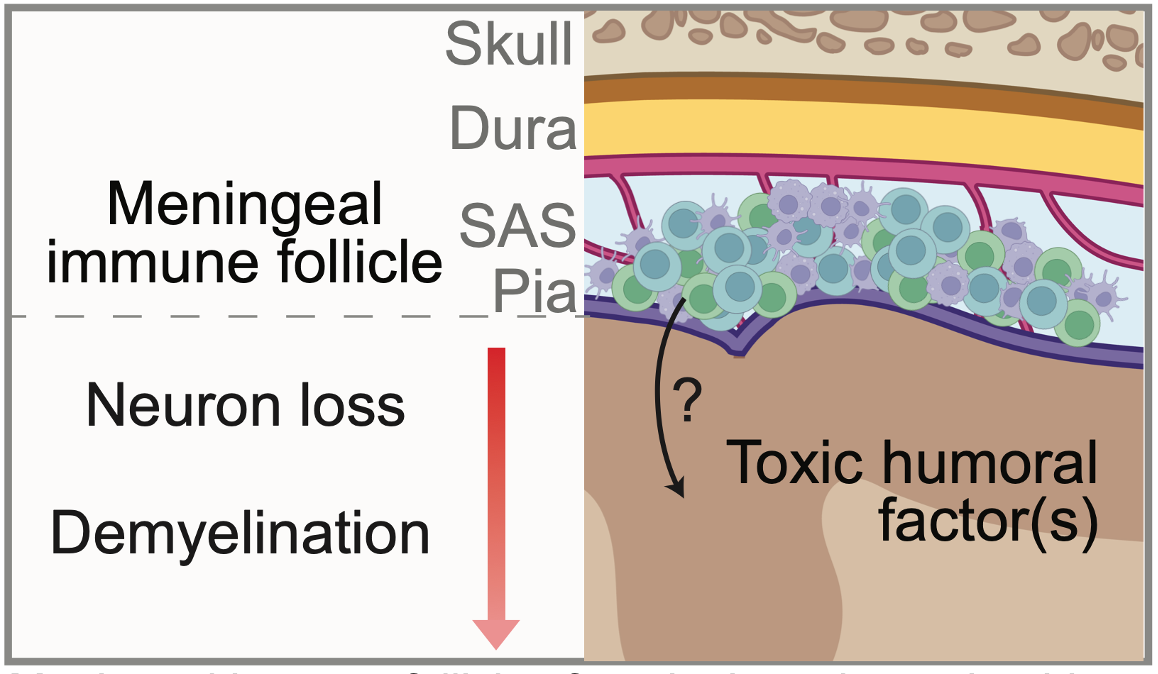
The brain is surrounded by a three-layer structure called the meninges, composed of the dura, arachnoid, and pia mater. Between the arachnoid and pia mater is the subarachnoid space, which houses cerebrospinal fluid. In multiple sclerosis, collections of immune cells called meningeal lymphoid aggregates (MLAs) develop in the SAS and are a local source of inflammatory molecules. Interestingly, grey matter lesions typically form under MLAs, and there is a gradient of increased cell loss towards to the surface of the brain. Grey matter pathology also occurs without local infiltration of peripheral immune cells into the brain parenchyma. Collectively, these observations suggest that humoral factor(s) originating in MLAs enter the brain and mediate grey matter pathology, but the molecule(s) themselves are unknown. We are using spatial transcriptomics to help address this question.
Gadani SP*, Singh S*, Kim S, Smith MD, Calabresi PA, Bhargava P. Spatial transcriptomics of meningeal inflammation reveals variable penetrance of inflammatory gene signatures into adjacent brain parenchyma. eLife. 2023. * Co-first author.